
INTRAVENOUS UROGRAPHY
The urinary system, also known as the renal system or urinary tract, consists of the kidneys, ureters, bladder, and urethra. The purpose of the urinary system is to eliminate waste from the body, regulate blood volume and blood pressure, control levels of electrolytes and metabolites, and regulate blood ph. The urinary tract is the body's drainage system for the eventual removal of urine.
The kidneys have an extensive blood supply via the renal arteries which leave the kidneys via the renal vein. Each kidney consists of functional units called nephrons. Following filtration of blood and further processing, wastes (in the form of urine) exit the kidney via the ureters, tubes made of smooth muscle fibres that propel urine towards the urinary bladder, where it is stored and subsequently expelled from the body by urination (voiding). The female and male urinary system is very similar, differing only in the length of the urethra.
Urine is formed in the kidneys through the filtration of blood. The urine is then passed through the ureters to the bladder, where it is stored. During urination, the urine is passed from the bladder through the urethra to the outside of the body.800– 2,000 millilitres (mL) of urine are normally produced every day in a healthy human. This amount varies according to fluid intake and kidney function.

INTRAVENOUS UROGRAPHY EXAMINATION
Urography is a radiologic technique used for the evaluation of the genitourinary system—specifically, the kidneys, ureters, and bladder. Although originally performed using plain radiographic techniques, advanced imaging modalities have been progressively refined such that computed tomography (CT) and/or magnetic resonance imaging (MRI) have largely replaced excretory urography (EU) as the optimal way to image the genitourinary system.
Indications
Suspected urinary tract pathology.
Repeated infections -focus, damage
Hematuria
Investigation of hypertension not controlled by medication in young adults.
Renal colic.
Trauma.
Contraindication
General contraindications to contrast agents.
Diabetes,
Thyrotoxicosis
Pregnancy
Raised urea creatinine
Metformin therapy
Patient Preparation
Bowel preparation.
Basic psychological preparation with reassurance & explanation of the technique
Nothing by mouth (NBM) for 5 hrs.
The bladder should be emptied immediately before the exam.
Previous experience with iodinated contrast media.
Abdominal surgery, Allergies, drugs history.
Contrast
Inject nonionic contrast Or LOCM as a bolus, 30-60 sec
Adult 50-100 ml
Pediatric 1ml/kg

Adverse reaction of contrast
True contrast reactions are uncommon
Most commonly seen are minor side effects
Flushing
Metallic taste in my mouth
Tachycardia
Usually, resolve within a few minutes
TERMINOLOGY
Urogram
Visualization of kidney parenchyma,
Calyces and pelvis resulting from IV injection of contrast
Pyelogram
Describes retrograde studies visualizing only the collecting system So, IVP is a misnomer, it should be IVU
Cystography
Describes visualization of the bladder
Urethrography
Visualization of urethra
Cystourethrography
A combined study to visualise bladder and urethra
Film technique
Preliminarily films
Scout view
Additional views
Scout film for IVP
Supine, full-length AP abdomen, full inspiration
The lower border of the cassette at the level of the symphysis pubis, beam centred in the midline at the level of iliac crests
Often difficult to fit this large area on a single radiograph may need….
14 x 17 of the abdomen
10 x 12 of the lower pelvis
Purpose
Calcifications
Abnormal soft tissue
The air within the urinary tract
Bony abnormalities
Determine if a contraindication to abdominal compression exists
Additional views
Oblique Views (35-degree posterior oblique)
Good for questionable ureteral lesions
For differentiating extrinsic and intrinsic renal and ureteral masses
Visualization of the posterolateral aspect of the bladder
Supine AP - of renal areas in expiration
IVP STUDY
Routine Films for IVP
Immediate Film
AP of renal areas.
10-14 sec after injection
Shows Nephrogram
5 min Film
AP of renal areas
Determine if excretion symmetrical
Assess the need to modify the technique
Application of compression band balloon at the level of anterior superior iliac spines
Compression contraindicated in
Recent abdominal surgery
Renal trauma
Large abdominal mass or aortic aneurysm
Already dilated calyces on 5-min film
10 min Film
AP of renal areas
Compression is released if the pelvicalyceal system adequately visualized
Release film
Supine AP abdomen
Whole urinary tract
Empty bladder
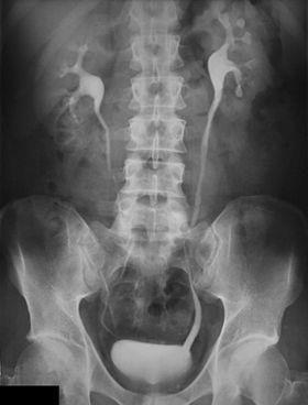
Post voiding Film
Full length OR
Coned view of the bladder (tube 15-degree caudal, centred 5 cm above symphysis pubis)
Must be obtained immediately after voiding
To determine residual urine in the bladder >, especially in older male patients
To look for bladder neoplasms
VUJ calculi
Urethral diverticulum in females
Additional views
Delayed Views
1 hour to 48 hours- in cases of obstruction
Better to CT patients for immediate diagnosis
Prone film
Helps fill ureteral areas not seen in the supine position since the upper ureters are more anterior than the kidney.
Erect film
Promotes emptying of collecting system.
Optimal for showing bladder hernias.
Shows layering of calculi in cysts.
Demarcates areas of ureteral obstruction better than prone views.

Hydronephrosis

IVP showing Grade IV hydronephrosis in the right kidney due to ureteropelvic junction obstruction

Bladder stone